- Undergraduate
Bachelor's Degrees
Bachelor of ArtsBachelor of EngineeringPartner School Dual-DegreeUndergraduate AdmissionsUndergraduate Experience
Quick Links
- Graduate
Doctoral Degrees
Doctor of PhilosophyPhD Innovation ProgramDoctor of Medicine-PhDGraduate AdmissionsGraduate Experience
Quick Links
- Research
Research by Program Area
Quick Links
All Thayer News
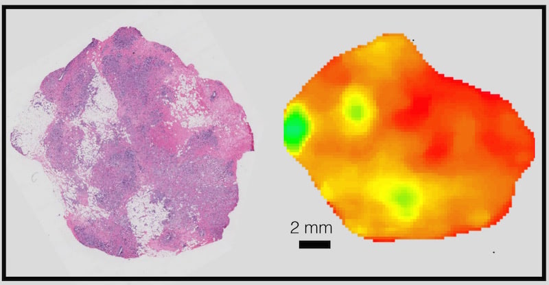
Left: Histology section of invasive carcinoma of the breast. Right: Optical property map where green = fat; orange-yellow = highly cellular invasive regions; and red = connective tissue with high collagen density.

David "Bo" McClatchy '13
New Wide-Field Imaging Technique Is One Step Closer to Helping Cancer Surgeons
Jun 15, 2016
A team of researchers from Dartmouth and the National Institute of Standards and Technology (NIST), with funding from both NIST and the National Institutes of Health (NIH), have published a new study on their method of quickly scanning large fields of view for microscopic-level details. The approach has wide-ranging potential applications including for surgical procedures.
"Doctors use a microscope to determine if tissue is normal, but during surgery, they can't use a microscope everywhere," says Stephen Kanick, Dartmouth engineering professor and a co-author of the study. "This approach lets the surgeon image the full field to show areas of abnormal microstructure and therefore show where to point the biopsy needle."
The study, entitled "Wide-field quantitative imaging of tissue microstructure using sub-diffuse spatial frequency domain imaging," was published in Optica, The Optical Society's journal for high-impact research in the field of optics.
Authors include researchers from the Optics in Medicine group at Dartmouth's Thayer School of Engineering, the Department of Pathology and Norris Cotton Cancer Center at Dartmouth-Hitchcock, and the Quantum Electromagnetics Division of NIST.
Left: Histology section of invasive carcinoma of the breast. Right: Optical property map where green = fat; orange-yellow = highly cellular invasive regions; and red = connective tissue with high collagen density.
This new imaging technique is sensitive to differences in microstructure — such as density and either particle or cell size as well as characteristics of the extracellular matrix — and can provide a visual representation, or map, of these differences over a wide field of view.
"It's like if you were using Google Earth and you wanted to determine which houses are actually clusters of condominiums," explains Kanick. "Our approach could tell you where to look for the condos without having to do the work of zooming in over and over again."
David "Bo" McClatchy '13
"We have a grant right now to investigate the effectiveness of combining this approach with CT imaging," added David "Bo" McClatchy '13, Dartmouth engineering PhD candidate and first author on the study. "This should enable a surgeon to see both the tumor core and also scan the surface of the resection for cancer cells." Because if there's cancer on the surface, then there might still be cancer left in the body.
"And in the future, we want to translate the optical properties data into maps that are even easier to interpret," says McClatchy. "Because as long as you know what you're looking for, the applications of this approach are wide-ranging." Applications include evaluating wound healing, and fast, effective characterization of food and materials.
Added Kanick, "There's also potential for incorporating this technology directly into surgical microscopes and endoscopes to guide doctors to problem spots in real time."
For contacts and other media information visit our Media Resources page.